ICSE Complete Study Guide: Cell DivisionMitosis, Meiosis & The Cell Cycle
Cell division is one of the highest-weightage topics in the ICSE Class 10 Biology syllabus — and one of the most misunderstood. Most students memorise definitions and diagram stages. Top scorers understand the why behind every stage. This guide covers the complete ICSE cell division syllabus: SA:V ratio, Karyoplasmic Index, DNA structure, the cell cycle, mitosis, meiosis, and examiner-approved diagram techniques.
Why Do Cells Divide? The Physics Behind It
A foundational ICSE exam question: why can’t a cell just keep growing?
Surface Area to Volume (SA:V) Ratio As a cell grows, its volume (s³) increases far faster than its surface area (6s²). The plasma membrane — the cell’s only interface for nutrient intake and waste removal — becomes overwhelmed. A smaller SA:V ratio means an inefficient cell. Division restores a high SA:V ratio, making each daughter cell metabolically effective again.
Karyoplasmic Index (KI) KI = Nuclear Volume ÷ Cytoplasmic Volume
The nucleus governs all cellular activity. When the cytoplasm expands too far, the nucleus can no longer manage it. A falling KI triggers the cell cycle, restoring the balance between nucleus and cytoplasm.
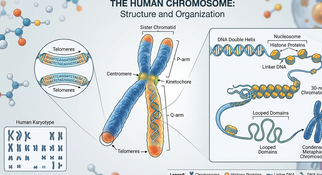
Discovered by Watson and Crick (using Rosalind Franklin’s X-ray data), the DNA double helix is built from nucleotides containing a phosphate group, deoxyribose sugar, and a nitrogenous base.
Base Pairing Rules:
- Adenine (A) pairs with Thymine (T) — 2 hydrogen bonds
- Cytosine (C) pairs with Guanine (G) — 3 hydrogen bonds
Memory tip: “AT the double, CG triple”
Purines (double ring): Adenine, Guanine | Pyrimidines (single ring): Cytosine, Thymine
DNA compacts by winding around histone proteins to form nucleosomes (“beads on a string”), which coil further into chromosomes visible during division.
The Cell Cycle: Interphase + M Phase
Interphase (often mislabelled “resting phase”) is a period of intense preparation:
- G₁ Phase: RNA, protein, and organelle synthesis; cell doubles in size
- S Phase: DNA replication — chromosome number stays the same, but DNA content doubles; each chromosome now has two identical sister chromatids
- G₂ Phase: Final checks on DNA integrity; spindle proteins synthesised
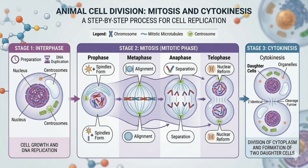
Mitosis: Equational Division (Somatic Cells)
Mitosis produces two genetically identical diploid (2n) daughter cells — essential for growth and tissue repair.
| Stage | Key Events |
|---|---|
| Prophase | Chromatin condenses; nuclear envelope breaks down; centrioles migrate (animal cells); spindle forms |
| Metaphase | Chromosomes maximally condensed and aligned at equatorial plate — best stage for counting |
| Anaphase | Centromeres split; sister chromatids pulled to opposite poles |
| Telophase | Nuclear membranes reform; chromosomes uncoil; nucleolus reappears |
Cytokinesis:
- Animal cells: Cleavage furrow pinches inward
- Plant cells: Cell plate grows outward from centre
ICSE Diagram Tip: Always draw and label the cleavage furrow (animal) or cell plate (plant). Examiners award specific marks for this.
Meiosis: Reduction Division (Reproductive Cells)
Meiosis occurs in the testes and ovaries and produces four genetically unique haploid (n) gametes through two successive divisions.
Meiosis I — Reductional Division
- Prophase I: Homologous chromosomes pair (synapsis), forming bivalents. Crossing over occurs at chiasmata — non-sister chromatids exchange DNA segments, creating genetic variation.
- Metaphase I: Bivalents (not individual chromosomes) align at the equatorial plate
- Anaphase I: Homologous chromosomes separate → chromosome number halved
- Telophase I: Two haploid cells form
Crossing over is the most important event in meiosis — it generates the genetic variation that drives evolution.
Meiosis II — Equational Division Similar to mitosis: sister chromatids separate, yielding four unique haploid cells. No DNA replication occurs between Meiosis I and II.
Mitosis vs. Meiosis: The ICSE Comparison Table
| Feature | Mitosis | Meiosis |
|---|---|---|
| Site | Somatic cells | Reproductive cells |
| Purpose | Growth and repair | Gamete formation |
| Daughter Cells | 2 (identical) | 4 (unique) |
| Chromosome No. | 2n → 2n | 2n → n |
| Crossing Over | Absent | Prophase I only |
| Genetic Outcome | Identical clones | Maximum variation |
ICSE Diagram Technique: How to Score Full Marks
- Use a sharp HB pencil — never pen or felt-tip
- Draw diagrams large enough to label clearly
- Use straight, horizontal label lines — no arrows, no crossing lines
Critical plant vs. animal cell distinction (most common mark-loss point):
- Plant cells: No centrioles; division via cell plate
- Animal cells: Centrioles and asters present; division via cleavage furrow
Quick Revision Checklist
✓ SA:V ratio decreases as cell grows → triggers division ✓ Karyoplasmic Index falls → nucleus loses control → division begins ✓ S phase = DNA replication | G₁/G₂ = growth and preparation ✓ Mitosis → 2 identical diploid cells | Meiosis → 4 unique haploid cells ✓ Crossing over occurs in Prophase I of Meiosis only ✓ Plant cells = cell plate | Animal cells = cleavage furrow
Summary
Cell division in the ICSE Class 10 Biology syllabus isn’t just memorisation — it’s physics, logic, and biological necessity. Cells divide because the SA:V ratio becomes unsustainable and the nucleus loses governance of its cytoplasm. Mitosis ensures genetic continuity for growth and repair. Meiosis ensures genetic diversity for evolution. Master the why behind each stage, practise labelled diagrams precisely, and use these comparison tables for last-night revision. That’s the most reliable path to full marks.